

ULTRASOUND & X-RAY
What is Radiography?
Radiography is the process of taking x-rays and interpreting the results. X-rays are a form of electromagnetic radiation that ultimately produce an image on a type of photographic film that can be interpreted to give an idea of physical changes. Most pets will require sedation or general anaesthesia to obtain diagnostic images as they are unlikely to keep still long enough to produce the image. As x-rays are a form of radiation, staff members performing the task must be appropriately protected with lead gowns, in a room with lead lined walls and their exposure levels are monitored routinely.
At CVC, we have top quality digital radiography, which allows us to obtain images much faster than with conventional radiography methods (development of the film in a dark room), enabling us to obtain better quality images, quicker than before, so enabling us to have a shorter, safer anaesthetic time for our patients.
Why does my pet need an x-ray?
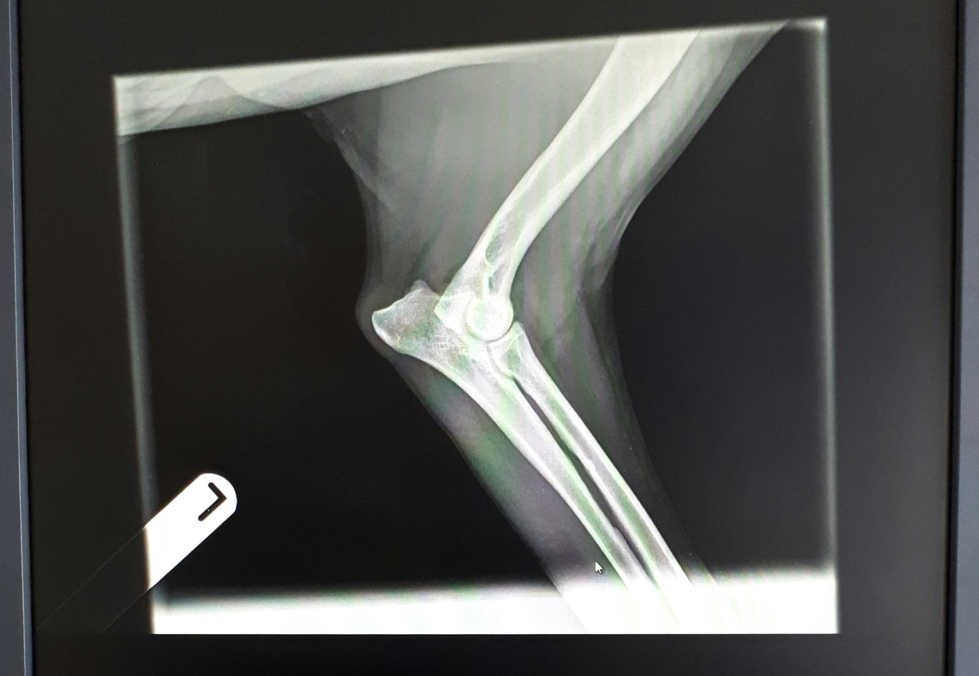
Our pets can need x-rays for a range of reasons. Principally we perform x-rays for bone disorders e.g. fractures/dislocations/or diagnosis of arthritis. However, x-rays can also be very useful for seeing soft tissue densities, e.g. organs or tumours so may be suggested in these circumstances. The use of x-rays for tumour diagnosis has been a little superseded by the use of ultrasound, but radiography still has a place. It can be useful for assessing any evidence of cancerous spread to other organs such as the lungs to assess prognosis. The main downside of this is that by the time spread is visible on radiography, the organs are already fairly heavily affected.
Ultrasonography
What is Ultrasonography?
Ultrasonography has become the second most commonly used imaging format in veterinary practice. It works through production of sound waves through a transducer, with the addition of ultrasound gel on the pet, to produce vibrations which bounce off the pets organs to produce an image on the screen for the vet to analyse. The sounds waves travel at different speeds through different tissues, giving us a variation in tissue densities to allow us to identify the various structures. Unlike radiography, ultrasound produces a dynamic image (one that moves in real time), so it is very useful for evaluating things like blood flow through organs and also for ultrasound guided sampling methods. The biggest downside of ultrasound is that it does not penetrate bones or gas, so this can affect the quality of images obtained.
Why does my pet needs ultrasonography?
We may advise ultrasound for your pet if there is any clinical suspicion of underlying disease. We mostly use it for evaluation of the abdominal organs, but increasingly it is being used for scanning of the heart (echocardiography), and muscles, tendons and ligaments. It allows us to establish most likely cause of a problem, thus give us a better chance to obtain diagnostic samples, to give you a better idea of prognosis for your little ones.